Erongo
Radiology
Erongo Radiology is a private Radiology facility committed to providing the highest quality patient care.

Erongo
Radiology
Erongo Radiology is a private Radiology facility committed to providing the highest quality patient care.
Erongo Radiology is a private X-ray facility committed to providing the highest quality patient care. Quality imaging, diagnostic excellence and the needs of our patients form the cornerstone of our service ethos. Our motto is: Quality care by people who care.
Being a multi-specialty Radiology Practice, our team of South African HPCSA certified and Allied Health Professions Council of Namibia registered, diagnostic Radiologists are committed to providing the highest quality radiological services possible to our patients through our state-of-the-art radiology equipment.
We have the following imaging modalities available at our main branch at the Welwitschia Hospital: General X-rays, Fluoroscopy, Ultrasound, Low Dose CT Scan, Mammography and MRI.
Computed Axial Tomography (CT Scan) is an X-ray powered technology using a scanner that produces cross sectional images of the body which can be viewed in two or three dimensions. These scans are used for examination of the brain, bone density, chest and abdomen and for fine detail examinations of the spine and joints.
Plain X-rays are the basic imaging method for diagnosing disease. Plain X-rays of the chest, sinuses, bones and spine are used as the primary method for diagnosing infections, trauma and diseases of bones due to wear and tear.
Magnetic Resonance Imaging (MRI) is a safe and painless test that uses magnetism and radio waves to produce detailed images of the body's organs and structures. Unlike CAT scans or X-rays, MRI doesn't use radiation.
Fluoroscopy is a type of medical imaging that shows a continuous, moving x-ray image on a monitor.
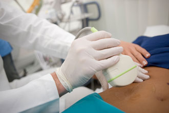
Ultrasound is a simple, safe and non-invasive method of evaluating the abdominal and pelvic organs. A routine abdominal study will examine the liver, gallbladder, kidneys, spleen and pancreas. Ultrasound is also useful for examining the aorta to exclude aneurysms. Ascites are readily detectable. Ultrasound has limited value in examining the intestine but may detect focal bowel wall thickening or bowel related masses.
This is an X-ray technique which uses very low dose X-rays for imaging of the breasts. All women over the age of 40 should have an annual mammogram. Ultrasound is used in many cases as an additional examination, to determine the nature of abnormalities detected on mammography or for added information in those women who have very dense breast tissue. There is no evidence that mammography itself is dangerous or can produce cancer.
Bone densitometry is used to assess the strength of bone. The tests are done with a CT scan machine. Bone densitometry is used for the diagnosis of osteoporosis and for monitoring its treatment.
| Dr J.M. Kabongo | M.D. MMED (RAD.D)(US), FCRAD (DIAG)(SA) |
| Dr L. Madisha | MBChB Radiology (Kwazulu-Natal), FC RAD (DIAG)(SA) |
© 2025 Blue Crane Web Development
